Physical Address
304 North Cardinal St.
Dorchester Center, MA 02124
Physical Address
304 North Cardinal St.
Dorchester Center, MA 02124
Exploring Cutting-Edge innovation in Medicine and Healthcare
Exploring Cutting-Edge innovation in Medicine and Healthcare

Tiny Maps, Huge Impact: Microscopic Maps Pave the Way for a Healthier Future.
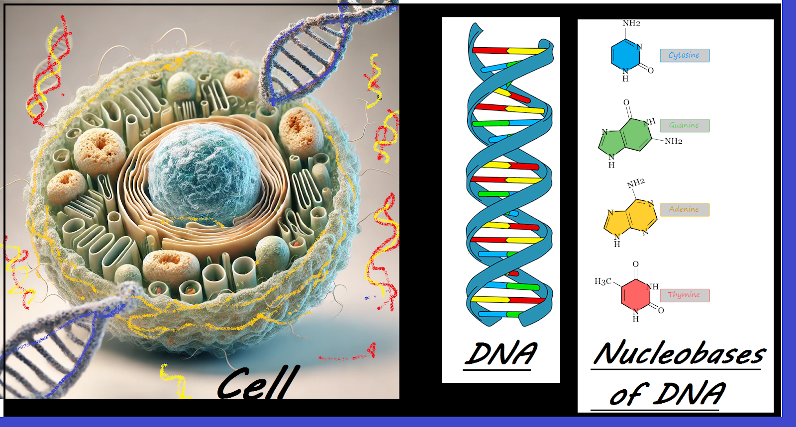
The cell, first discovered by Robert Hooke in 1665, represents the fundamental unit of life, functioning similarly to miniature computers that interact with their environment through biochemical processes. Cells utilize an array of proteins for decision-making and communication, determining actions such as proliferation and differentiation. The discipline of cell mapping aims to delineate how proteins collaborate in complexes to maintain cellular health, creating comprehensive maps of genetic and physical interactions within cells. Cell mapping aspires to establish a reference map of healthy human biology, facilitating improved treatment for various diseases, including cancer and diabetes. Additionally, this endeavor draws from genomics and molecular biology, focusing on cataloging diverse cell types across different life stages and understanding their evolution in relation to ancestry, geography, gender, and age.
The Human Genome Project (1990-2003) was a landmark scientific initiative that sequenced human DNA, fundamentally enhancing our understanding of genetics and advancing medical practices. The project’s achievement of creating the first comprehensive reference of the human genome has been pivotal in transforming health and disease understanding, driving significant advancements in global healthcare. Subsequently, the Human Cell Atlas (HCA) project was launched in 2016 as a major effort to map all human cell types throughout various stages of life. This international collaboration aims to document the cellular composition of healthy human tissues.
Dr Aviv Regev, co-chairwoman of HCA, explained mapping of cells:
“Cells are the basic unit of life, and when things go wrong, they go wrong with our cells, first and foremost. Once we have this map, there are many, many things that we are able to do. We are able to better find the causes of disease and understand how they operate. We need this map when we try to actually develop new medicine. And if we don’t know what the cells look like and what the healthy state looks like, we cannot actually revert a disease cell into a healthy cell.”
Professor Sarah Teichmann, co-chairwoman of the HCA, also explains:
“This is a major milestone that marks a great leap in our understanding of the human body. By creating a comprehensive reference map of the healthy human body – a kind of ‘Google Maps’ for cell biology – it establishes a benchmark for detecting and understanding the changes that underlie health and disease. This new level of insight into the specific genes, mechanisms and cell types within tissues is laying the groundwork for more precise diagnostics, innovative drug discovery and advanced regenerative medicine approaches.”
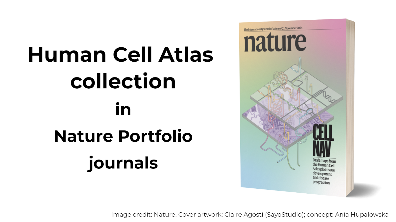
Researchers from all over the world are advancing the use of innovative technologies, including single cell transcriptomics, spatial genomics, and computational techniques, to study millions of cells from more than 10,000 individuals globally. These efforts have led to significant findings, including insights into placenta and skeleton formation, brain maturation, and lung responses to Covid-19. Approximately 3,600 HCA members have collaborated on this monumental task, with results disseminated through the journal “Nature.” The research methodology focuses on three core principles: measuring protein interactions under varying conditions, conducting genetic studies to observe cell behavior in the absence of specific genes, and integrating physical and genetic maps to better understand cellular functions and their implications. Although, Mapping every cell in the human body seems impractical, researchers estimated there are around 37.2 trillion in an adult, the DNA within each cell constitutes the genome, and nearly every cell contains a complete copy of it. When the genomes of two individuals are compared, they exhibit a 99.8–99.9% similarity; it’s the 0.1- 0.2% variation in our genome that interests the researchers to focus on understanding these genomic variations that are helpful in the prediction, prevention, diagnosis, and treatment of diseases.
“Single-cell transcriptome sequencing” has enabled the mapping of individual cells and the compilation of an atlas by examining the messenger molecules that convey genetic instructions from a cell’s nucleus to its protein synthesis machinery. These messengers are made by transcribing genes into a DNA-like chemical called RNA. Fluorescent chemicals attached to molecular tags that will stick only to particular RNA messengers show up the cells containing those messengers. This technology enhances the understanding of anatomical details by providing a zoomable view, akin to navigating an internet map of the Earth. Shannon Hughes from America’s National Cancer Institute highlights the significance of transcriptome sequencing in skin cancer research, through the creation of the Human Tumor Atlas. Tumors, with their genetic mutations, are well-suited for this analysis, Dr. Hughes and her team have identified pre-cancerous skin cells that do not possess the full complement of mutations necessary for cancer progression. They have noted that these cells attract T-lymphocytes—immune cells that penetrate the skin tissue to address potential threats.
A cell atlas of the human gut encompasses a comprehensive map of the gut, extending from the mouth to the anus, examining various cell types, locations, and interactions with neighboring cells, detailing both healthy and diseased tissues. This resource is significant for exploring and treating conditions such as ulcerative colitis and Crohn’s disease. Out of 1.6 million cells analyzed, gut metaplastic cells were identified, which appears to contribute to increased inflammation in individuals affected by inflammatory bowel diseases.
Researchers from the National Heart and Lung Institute at Imperial College London and the Cambridge Stem Cell Institute have successfully identified and mapped various types of blood vessels in the human body for the first time. This knowledge is anticipated to facilitate the development of targeted treatments and addressing conditions such as diabetes, and other inflammatory diseases. Co-first author, Sam Barnett at the National Heart and Lung Institute, London, said:
“Now, we have a more complete understanding of the molecular and cellular architecture of vessels, and that could allow the creation of drugs that precisely target specific vascular cells in diseased organs and tissues. For example, if scientists want to make a drug that targets blood vessels in the brain, they can now distinguish those cells from other organs’ vessels, enabling them to produce a medication that only affects the intended ones and reduces unwanted side-effects.”
In a recent study published in the journal “Nature,” have developed a comprehensive blueprint detailing human embryonic skeletal development. Notable findings include the role of cartilage as a scaffold for bone development, the identification of a distinct subpopulation of human skeletal stem cells, and a detailed lineage map showing the transformation of these stem cells into various skeletal tissues. The insights from this research are expected to enhance understanding of conditions like osteoarthritis. Additionally, the researchers propose that this resource could be instrumental in assessing the impact of existing or future medications on skeletal growth during pregnancy.
During the Covid pandemic, the Human Cell Atlas played a crucial role by providing detailed maps of the human body, which helped scientists predict the virus’s movement and identify the nose, mouth, and eyes as primary entry points. This cell mapping also revealed the activity levels of various immune cells in severe cases, significantly impacting the development of vaccines and therapies. Furthermore, recent discoveries from the global HCA consortium have pinpointed specific cells in the nose and eyes that may facilitate the transmission of the SARS-CoV-2 virus.
Recent advancements in cell mapping have led to the identification of tuft cells, a rare cell type within the lungs, which helped in understanding various respiratory diseases. These chemosensory cells, located in the epithelial lining of the lungs and intestines—also referred to as brush cells within the respiratory epithelium—play significant roles in inflammation and dysplasia, potentially contributing to conditions like asthma and chronic rhinosinusitis.
A project focused on mapping the cells of the endometrium, which is the mucosal lining of the uterus, is underway. Its goal is to gain insights into the physiology of the endometrium and to identify the factors that contribute to diseases affecting this tissue.
The mapping of cells is a complex and costly endeavor due to the need for advanced technologies such as single-cell sequencing, computational resources, and specialized personnel. Nevertheless, advancements in technology are gradually lowering these costs. Researchers aim to create a preliminary Human Cell Atlas by 2026, which will expand to encompass billions of cells from various organs and tissues. This collaborative global initiative involves notable institutions including the Wellcome Sanger Institute and Cambridge University (UK), the Broad Institute and NIH (USA), the Karolinska Institute (Sweden), the Max Planck Institute (Germany), the Beijing Genomics Institute (China), and the CSIR-Institute of Genomics and Integrative Biology (India).
Mapping of cells is essential for the progress of medical research, biotechnology, and personalized medicine. By gaining insights into cellular structures, functions, and interactions, researchers can create targeted treatments for diseases, advance regenerative medicine, and enhance drug discovery. Additionally, it supports developments in artificial intelligence and bioinformatics, leading to further innovations in healthcare and the life sciences. As technological capabilities improve, the precision of cellular mapping will continue to transform medicine and human health.
————————————————————————————————–
Bibliography
This essay acknowledges the contributions of Google, Wikipedia, and www.independent.co.uk, https://www.humancellatlas.org, www.sanger.ac.uk and various websites appear during google search, that provided valuable information for its composition.

A closer look at the historical development:
St. Bartholomew’s Hospital (London):
Established in 1123 by Rahere, it began as part of the Priory of St. Bartholomew and later evolved into an independent institution. Today, it stands as one of the world’s oldest hospitals still in operation.
Byzantine Empire:
Around 370 AD, St. Basil the Great established a complex in Cappadocia (in present-day Turkey) that included a hospital. Similar institutions later spread throughout the Eastern Roman Empire.
Persian Empire:
In the 3rd century AD, the city of Gundeshapur (in modern Iran) was home to a renowned hospital and medical teaching center.
Ancient Greece:
Temples known as Asclepeia, dedicated to the healing god Asclepius, provided care but are generally not viewed as true hospitals by modern standards.
Ancient Rome:
While public hospitals were rare, the Romans did establish valetudinaria, hospitals for soldiers and slaves within military camps. Wealthy Romans sometimes also maintained private facilities on their estates.